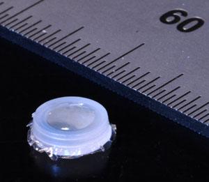
It looks like a tiny white breath mint. As Michael Cima, from the Massachusetts Institute of Technology says “With this, we are going to bring the laboratory into the patient”.
The little capsule is in fact an innovative tool in the struggle against cancer that can track the growth of a tumor without repeated invasive procedures.
This innovative tool is small enough to fit inside a needle and implant in the body during a biopsy. Magnetic nanoparticles fill the capsule’s hollow interior, each sporting a few monoclonal antibodies. These are proteins engineered to bind to molecules of interest, such as human chorionic gonadotrophin (hCG), a hormone that tumor cells overproduce in testicular and ovarian cancers.
A semi-permeable membrane allows molecules to flow into the capsule, but prevents the nanoparticles from drifting out. To read the sensor and evaluate whether a nearby tumor is receding or growing, doctors need only use an MRI scan to detect clusters of cancer-related molecules within the innovative tool.
Cima’s team tested the device in mice injected with human cancer cells. An MRI showed that the resulting tumors were increasing in size. In another study with mice, Cima transformed the tumor-monitoring device into a heart-attack detector by lining the inside of the capsule with antibodies that bind to three different proteins released by heart muscle cells when they burst open.
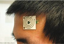
After embedding the sensor in the rodents’ skin and inducing the heart attack, Cima was able to precisely measure the severity of damage: the more protein accumulated in the monitor, the worse the heart attack and the stronger the MRI signal. Such a sensor device would be especially useful for people who experienced silent heart attack – which usually present little pain or obvious symptoms – as well as those at risk of a second heart attack.
Cima is now developing a more advanced prototype of the device that produces a signal simply by waving a magnetic wand over the embedded sensor.
Radiologist Heike Daldrup-Link at Stanford University in California is impressed by the prospect of tumor-trackers that can be read with handheld scanners, which could eliminate expensive MRI scans.
She also points out that current imaging techniques can have difficulty distinguishing tumor re-growth from healthy tissue that is reacting adversely to chemotherapy or radiation. “I think these devices could be very powerful, very useful,” she says.


